Musculoskeletal Imaging
Bringing adaptive image quality to point-of-care bone ultrasound. From bone probability maps and signal processing to deep learning beamformers that run in real time on handheld devices.
Bone Probability Maps
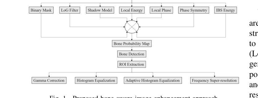
Bone probability maps (BPMs) are the connecting thread across our MSK imaging research. A BPM assigns every pixel in an ultrasound image a probability of belonging to a bone structure, built from local phase, feature symmetry, acoustic shadow detection, and integrated backscatter (IBS) energy.
In the earliest work, BPMs were combined with image enhancement techniques applied selectively to the region of interest above the detected bone surface. Gamma correction, histogram equalization, adaptive histogram equalization, and a frequency-based super-resolution method were evaluated on 22 sets of fundamental and harmonic shoulder ultrasound images.
The BPM has since become an attention mechanism inside deep learning architectures, guiding networks to focus on clinically relevant bony structures while suppressing irrelevant soft-tissue regions.
- FeaturesLocal phase, feature symmetry, shadow map, IBS energy
- DetectionGradient-based bone pixel identification with cubic spline fitting
- ROIMean tendon thickness ~5.2 mm above detected bone contour
- EnhancementAdaptive histogram equalization (PSNR 21.16, MSE 0.0078)
From RF Data to Clinical Images in Milliseconds
Three PatchGAN-based architectures that learn the beamforming process directly from raw radio frequency channel data, replacing computationally expensive algorithms with networks that run in under 5 milliseconds on a single GPU.
PatchGAN → DAS Accelerator
A U-Net generator with PatchGAN discriminator that reconstructs conventional DAS B-mode images directly from single plane wave RF data, learning both time-of-flight correction and apodization. Achieves image quality comparable to conventional DAS on in-vivo wrist datasets.
DOI →PatchGAN → MVDR Accelerator
Extends the PatchGAN framework to reconstruct multiple plane wave MVDR beamformed images from single plane wave RF data. Produces MVDR-quality images in 0.004 seconds, a 99.7% reduction from the 148 seconds required by conventional MVDR processing.
DOI →POCUS-Net
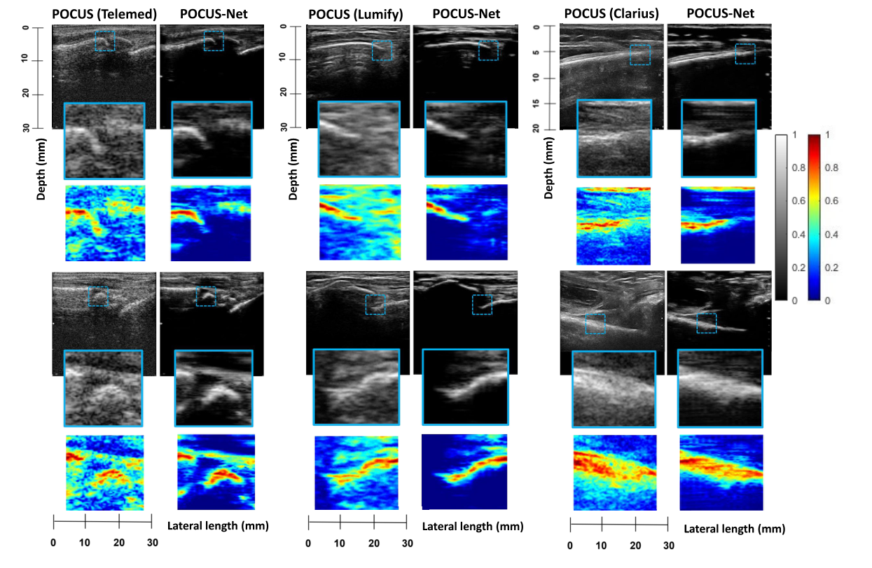
The flagship framework: a PatchGAN with IBS-derived intensity map conditioning that transforms DASB images into MVDR-quality outputs without requiring RF data. Validated on 1500 clinical pediatric wrist scans from three different POCUS probes (Telemed, Lumify, Clarius).
DOI →POCUS-Net: Pediatric Bone Visualization
POCUS-Net addresses the core limitation of handheld ultrasound for MSK assessment: POCUS devices produce images with poor contrast, high speckle noise, and limited resolution around bony structures because they rely on computationally simple delay-and-sum beamforming.
The framework transforms these low-quality DASB images into outputs that match the fidelity of computationally expensive MVDR beamforming, without needing access to the raw RF data that most commercial devices do not expose. An integrated backscatter (IBS) intensity map conditions the PatchGAN discriminator, acting as an implicit attention mechanism that focuses the network on bone structures.
Training used 1900 in-vivo wrist datasets from six adult volunteers and 1200 synthetic datasets generated from 20 pediatric subjects. Testing was performed on 1500 clinical scans from 24 children imaged with three different handheld probes. Compared to the input DASB images, POCUS-Net improved contrast ratio by up to 40%, signal-to-noise ratio by over 200%, and contrast-to-noise ratio by nearly 130%.
- ArchitectureU-Net generator (3 enc + 3 dec) with PatchGAN discriminator
- Training Data1900 in-vivo adult + 1200 synthetic pediatric wrist datasets
- Test ProbesTelemed L15-7H40-A5, Philips Lumify L12-4, Clarius L15 HD3
- Subjects24 pediatric patients (normal and fractured wrists)
- ConditioningIBS-derived intensity map as discriminator attention input
- TextureGLRLM + GLCM analysis confirming bone microstructure preservation
From Image Enhancement to Diagnosis
Bone probability maps and domain adaptation techniques enable two clinical AI applications: automated fracture detection from 3D ultrasound and cross-device image translation for hip dysplasia screening.
Wrist Fracture Detection from 3D Ultrasound
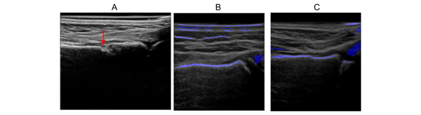
Bone probability maps computed from each frame of a 3D ultrasound sweep are vectorized and processed by recurrent neural networks (RNN, LSTM, GRU) to classify the presence of distal radius fractures. Validated on 30 pediatric 3DUS volumes (ages 0 to 17) scanned with a Philips iU22 at 13 MHz. The GRU model achieved 87% accuracy with the best balance of sensitivity (86%) and specificity (87%), outperforming CNN baselines including AlexNet, VGG-16, ResNet-50, and Inception V3 while using orders of magnitude fewer parameters (~0.5M vs. ~138M for VGG-16).
Domain-Aware Hip Ultrasound Translation
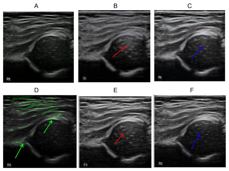
A domain-aware contrastive unpaired translation (D-CUT) framework that converts images from handheld Philips Lumify probes into pseudo-3DUS images matching the quality of conventional Philips iU22 probes. A bone probability map embedded in the GAN loss guides the discriminator to preserve structural fidelity around the acetabulum, ilium, and femoral head. Tested on 575 infant hip images from two clinical sites (University of Alberta Hospital and Westview Clinic) for developmental dysplasia of the hip (DDH) screening.
Related Projects
MSK imaging builds on and feeds into several parallel research threads across our group, from neural beamforming architectures to simulation infrastructure and structural signal processing.
The PatchGAN accelerators developed for MSK imaging share architectural DNA with the broader neural beamforming program that targets general-purpose adaptive beamformer learning.
View project → SimulationPOCUS-Net training used 1200 synthetic datasets generated with the SIMUS toolbox from clinical pediatric wrist images, extending the simulation infrastructure to MSK applications.
View project → Signal ProcessingBone probability maps build on the same local phase and feature symmetry analysis used in structural beamforming for characterizing specular vs. diffuse reflection.
View project → SystemsThe fracture detection work uses 3DUS sweep volumes acquired at the Stollery Children’s Hospital, connecting MSK clinical AI with the volumetric imaging pipeline.
View project → ClinicalReal-time muscle imaging for gesture recognition and prosthetic control shares the MSK domain, with both efforts targeting skeletal structures visible in ultrasound.
View project → Signal ProcessingNeedle guidance in MSK interventions benefits from the same bone-aware image enhancement, as improved bone visualization helps localize injection targets near bony landmarks.
View project →