LiveDemos
Interactive demonstrations of methods developed in our group. Each demo runs the trained model on real or simulated ultrasound channel data, so that the effect of the method can be seen directly, together with the corresponding publication.
PIVOT: Physics-Informed Variable Objective Tuning
Single plane wave beamforming where the balance between spatial resolution and contrast is selected by the user with one slider, using one trained network.
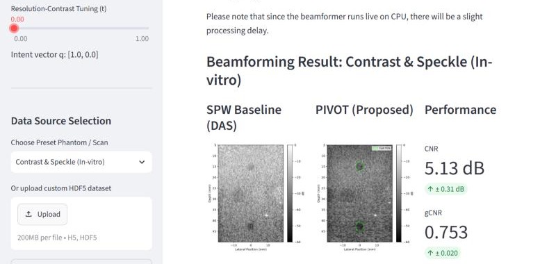
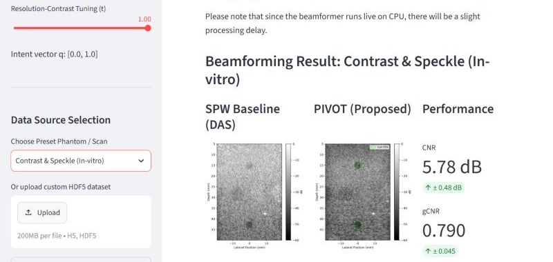
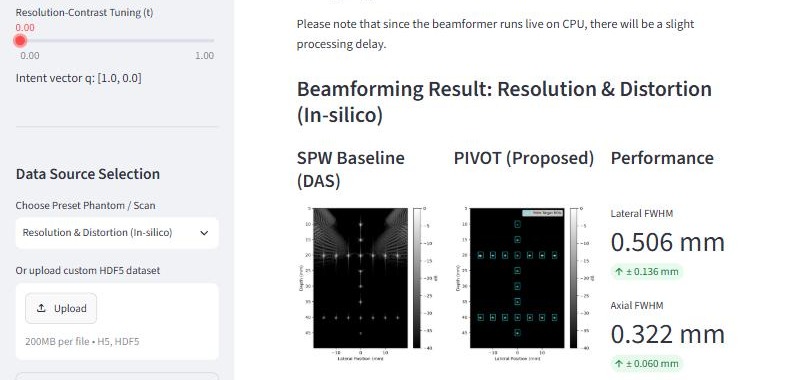
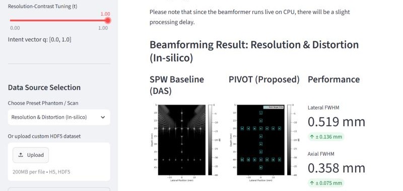
One slider between resolution and contrast
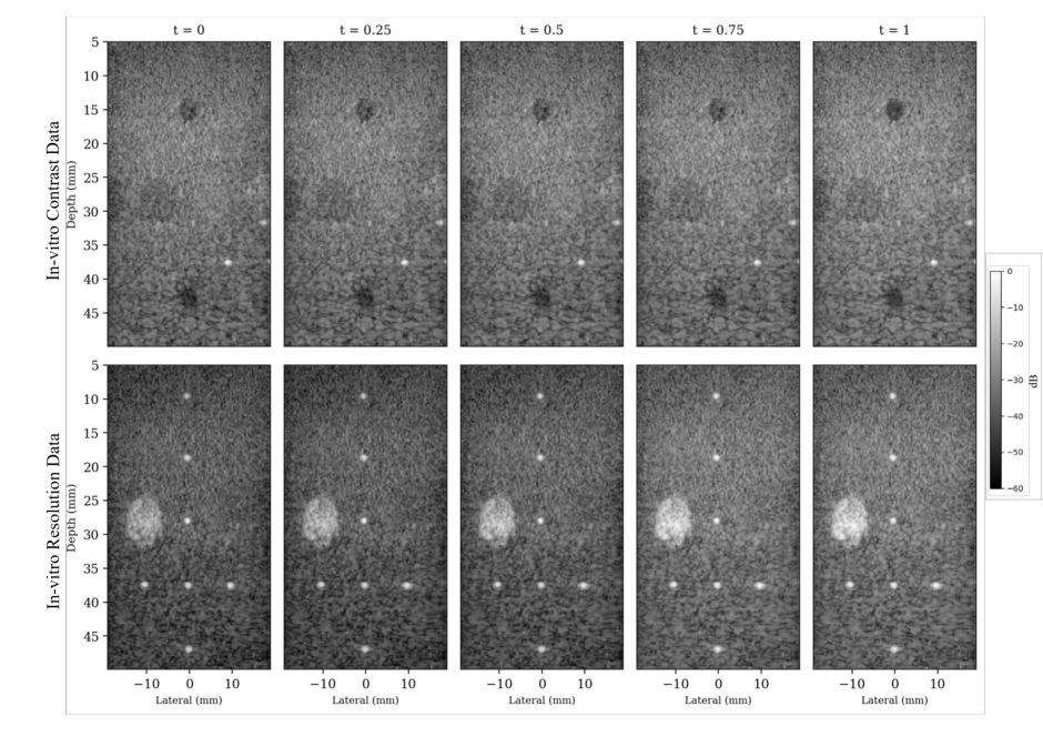
The slider sets the tuning value t between 0 and 1, which forms the intent vector q = [1 − t, t]. At t = 0 the network is conditioned for spatial resolution and at t = 1 for lesion contrast, with intermediate values giving operating points between the two. Each change of t re-runs the beamformer and updates the B-mode image and the quality metrics, shown beside the single plane wave delay and sum (DAS) baseline.
Six preset datasets are available, i.e., resolution and distortion, contrast and speckle (in-silico and in-vitro each), and carotid cross-sectional and longitudinal (in-vivo). A custom dataset can also be uploaded as an HDF5 file of up to 200 MB, and the required schema is given in the demo. The beamformer runs on CPU, hence each update takes a few seconds.
The captures on the left were taken at both ends of the slider. On the contrast phantom CNR increases from 5.13 dB to 5.78 dB when moving from t = 0 to t = 1, while on the point targets lateral FWHM increases from 0.506 mm to 0.519 mm, which is the expected resolution-contrast trade-off.
- ControlTuning value t from 0 (resolution) to 1 (contrast); q = [1 − t, t]
- PresetsIn-silico, in-vitro and in-vivo carotid datasets
- MetricsLateral and axial FWHM for point targets; CNR and gCNR for cysts
- Custom dataHDF5 upload, up to 200 MB per file
Opens in a new tab. The demo is hosted separately and runs on CPU.
Objective-conditioned receive apodization
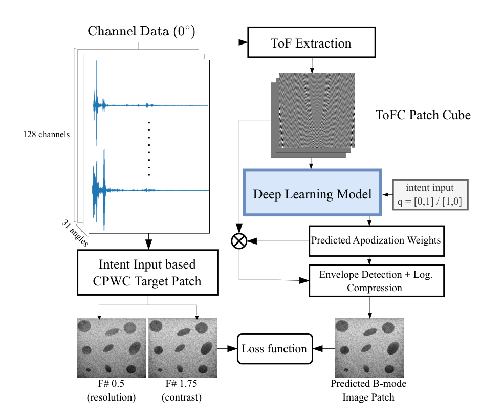
Plane wave imaging enables ultrafast acquisition, however the achievable resolution and contrast depend on the receive aperture, and conventional beamformers operate at a fixed aperture or F-number. Towards this, a physics-grounded, objective-conditioned deep beamformer is proposed, which predicts spatially varying receive apodization weights from single plane wave channel data. The objective is introduced through feature-wise linear modulation (FiLM), and each objective is tied to a physically defined receive aperture and supervised with the corresponding coherent plane wave compounding target.
The model is trained only on simulated channel data and evaluated zero-shot on the PICMUS and CUBDL datasets, including unseen probe geometries. On PICMUS it improves gCNR by up to 16.8% and axial resolution by 22% compared with the reference beamforming method, and switching between the objectives changes lesion contrast by up to 4.9% and lateral resolution by up to 11.4%.
- InputSingle unsteered (0°) plane wave, 128 channels
- ConditioningFiLM on objective vector q: [1, 0] resolution, [0, 1] contrast
- TrainingSIMUS (MUST toolbox) simulations, 350 acquisitions, 3,150 patches
- EvaluationZero-shot on PICMUS and CUBDL, including unseen probe geometries
- vs learned baselinesLateral FWHM reduced by 5.9%, gCNR improved by 6.6%
diffUS: Differentiable Ultrasound Co-design
Optimising the imaging system by gradient descent
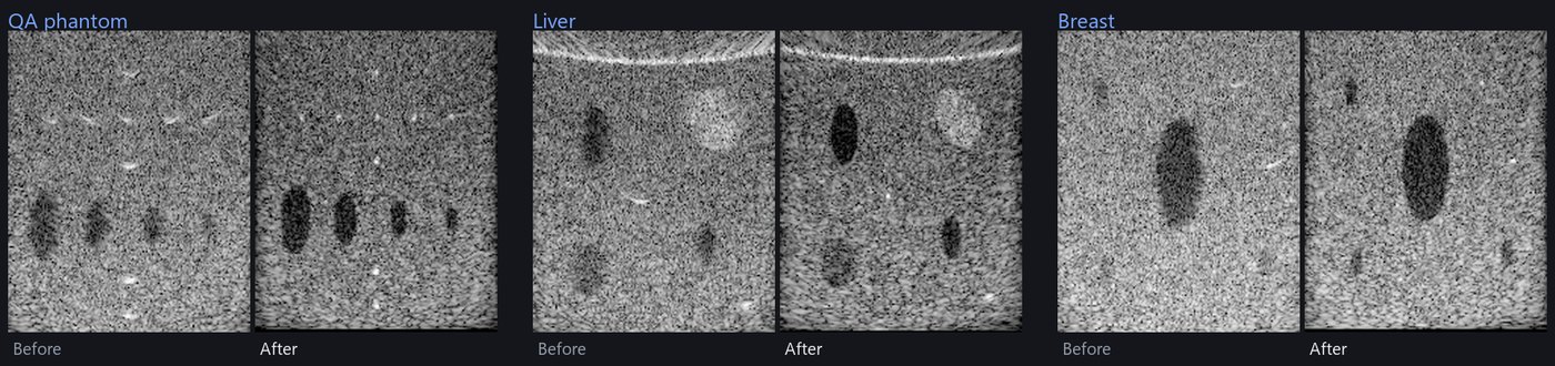
A JAX based differentiable simulator and beamformer, where the speed of sound and the transmit and receive apodization are optimised by gradient descent on image quality metrics, e.g., gCNR. Scene presets (QA phantom, liver, breast) are available, and an uploaded image can also be used as the scene.
Neural Beamforming at Pulse Echo
PIVOT is the latest work in our neural beamforming programme, which started in 2021 with CNN approximations of minimum variance beamforming.